Introduction to Soft Tissue Neck Xray
As a result of improvements in technology, soft tissue neck xray are improving over time. They have great importance in diagnosing many conditions, like infections or cancerous growths on the neck. As we look toward 2025, there’s so much to be excited about, new Technology—particularly how it will assist healthcare workers in solving emerging challenges! Technological advancement is vital for overall patient management and can help clinicians provide better care and improve diagnostic accuracy.
Together, we will study soft tissue neck xray, its importance for general patient management, and specific common gaps in interpretive accuracy radiologists encounter during the interpretation of the images. There’s much ahead of us regarding soft tissue nec imaging, let’s take this journey together!
Advancements in Technology: How Xray Imaging has Evolved
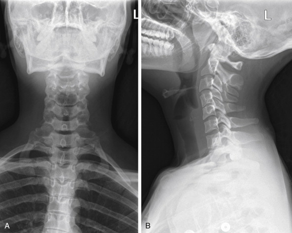
Over the years, soft tissue neck xray imaging has changed a lot. The use of traditional films has almost completely been replaced by digital radiography due to its increased speed and superior image quality. Compared to traditional devices, soft tissues can be better visualized within the neck for clearer images through digital technology with decreased radiation exposure. This improves diagnostic accuracy. Dedicated auxiliary software interprets images, aiding radiologists with their work. Important features that may be simple to miss are flagged by systems based on artificial intelligence tools, enhancing workflow and improving patient care.
Additionally, portable X-ray machines have begun to make an impact in emergency settings as they provide immediate evaluation without extensive mobility for patients, ensuring timely assistance. An additional pair of 3D imaging techniques is considered game-changing as well. They allow reconstruction and observation of important structures from various angles, improving assessment and treatment planning concerning complex cases involving the soft tissue neck area.
Challenges in Soft Tissue Neck Xray Interpretation
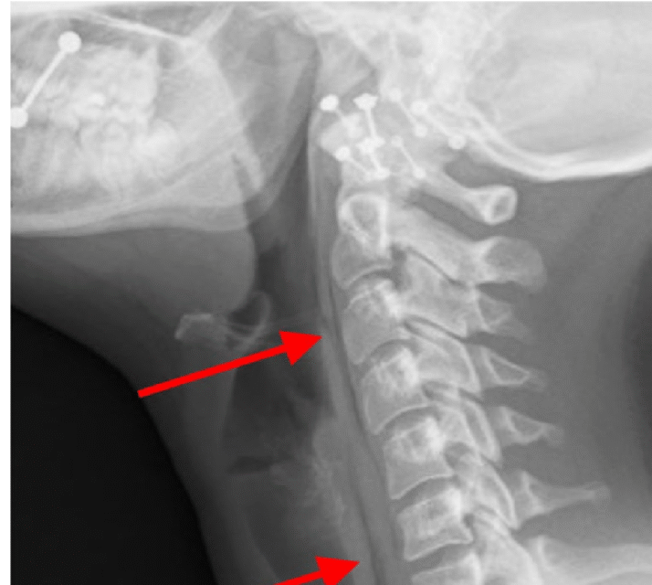
Soft tissue neck xray has many difficulties. Notable among these are overlapping structures, which obscure important features. Less experienced radiologists are particularly prone to the region’s complexities.
Every diagnostic process must result in distinguishing normal from abnormal changes. In detecting early infections or tumours, soft tissues’ subtle changes may hide them and blunt them.
Change in patient position might help to improve certain technical traits, but it can also add complications to an already challenging interpretative task, such as alignments shift, angles, and distorted images. Moreover, dental artefacts or other unrelated body parts can conceal basal vital pathology changes in transverse filters. All of this requires a lot of experience for proper imaging evaluation.
Solutions and Strategies for Overcoming Common Challenges
Using a systematic approach and soft tissue neck xray go hand in hand. In radiology, creating definable boundaries aids in consistent interpretation across various systems. These policies make certain that all relevant anatomy is included and assessed. Rest assured, multidisciplinary collaboration boosts the accuracy of healthcare diagnostics. A culture of active learning is created when there are regular case critiques or interdisciplinary meetings. This strengthens learners’ skills regarding interpretations and helps them feel confident about their decisions.
With the addition of traditional X-ray imaging, other methods like Ultrasound and CT scans can greatly aid with soft tissue neck pathologies as these fulfil a greater purpose when complemented by each other. For performing such roles, motivation with regard to continuous education is mandatory for radiologists. New standards or techniques emerging from attending workshops, as well as online courses, enhance their knowledge regarding practices scoped for imaging and strategies related to interpretation.
The Role of Radiologists in Soft Tissue Neck Xray Interpretation
Radiologists are essential professionals who interpret soft tissue neck xray as they each have unique skills and experience that allow them to spot minute details others may not see.
These physicians have extensive knowledge regarding anatomy and pathology, which helps them assess different conditions. They possess the ability to tell normal anatomical structures from pathological changes with great precision. Cooperation is very important for this discipline. Alongside other medical practitioners, radiologists also participate in taking care of the people to make sure all aspects are dealt with fully.
Their input greatly aids a person in accurately identifying an infection, tumour, or even trauma to the patient. Gaining more knowledge makes them better imaging interpreters, as well as advances in techniques. This ensures that radiologists remain up to date on everything technology-related and have sound clinched knowledge when it comes to work. Aside from being interpreters of ultrasound results, these specialists are active members of healthcare teams who discuss treatment plans based on findings for patients suffering from various neck conditions, which calls for a multidimensional approach to problem-solving.
Future Possibilities and Innovations in Soft Tissue Neck Xray
The future of soft tissue neck xray imaging is on the brink of remarkable improvements. Labelled as a remarkable radiographic exam with versatile applications, its optimization within the context of other techniques is indispensable. Radiologists’ workloads can benefit from automation since computer-assisted diagnostic systems powered by AI and machine learning can bring unparalleled precision to diagnostics. Routine checks have the potential to overlook critical details. These diagnostic tools can help radiologists by warning them of critical anomalies that require immediate attention.
In addition, computerized axial tomography (CAT) scans provide a series and slice imaging techniques with x-ray utilization for soft tissue structure imaging to aid in diagnosis and treatment planning improvement visually. In addition, remote* garner access to high-quality portable X-ray devices offered at great value. These sophisticated gadgets come equipped with potent imaging portals that can yield comprehensive images from difficult clinical settings. The immediate access they offer greatly improves emergency services provided in rural places or those known to lack fundamental infrastructure, in dire need of avenues.
Conclusion: The Importance of Regular Training and Education for Radiologists
This stream of innovation also requires continued education among radiologists. The subordinate and advanced level techniques and tools available for specialist interpretation require ongoing further education programs alongside surgical workshops. While interpreting soft tissue neck xray, continuous training courses enhance personal skills for their designated roles in healthcare teams. Additionally, working side by side with other medical specialists improves not only personal skills but overall common knowledge, leading to advancement in teamwork that’s required when dealing with patients.
With every new imaging method, there comes a need for adaptation, which serves as an obstacle course ready to be optimally solved whenever needed. Radiologist educators utilize challenges that enable them to take pride while offering specialized attention directed towards knowing how to maintain proper health care services for all patients. Creating and enforcing the atmosphere of endless learning contributes toward saying the crowning authority has been introduced into practice during all procedures performed in disciplines such as diagnosing medically advanced complex conditions, like those under the delicate tissues situated deep within what rounds neck regions would imply.